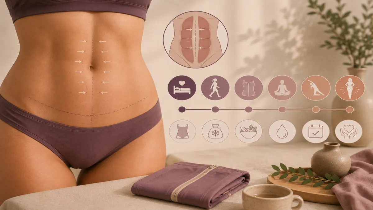
Before-and-after photos of diastasis recti surgery are among the most misleading images on the internet — not because they are fake, but because they almost never state when the "after" was taken. A photo at 3 weeks post-op looks radically different from one at 6 months, and the difference has nothing to do with technique. It is swelling, healing, and tissue maturation. This guide walks through what diastasis recti repair actually looks like at each recovery stage, what the surgery can and cannot achieve visually, and how to evaluate before-and-after galleries with a critical eye. For the technical overview of surgical technique, candidacy, and open versus laparoscopic approaches, see our diastasis recti surgical repair cluster page. Our broader muscle repair guide covers all plication options.
What Diastasis Recti Repair Actually Changes Visually
Diastasis recti is a separation of the left and right rectus abdominis muscles along the linea alba. The hallmark visual sign is a midline bulge — a dome or ridge that appears when you engage your core, and a soft, protruding lower abdomen even at rest. Surgical repair (plication) sutures the two muscle edges back together, restoring the midline tension of the abdominal wall.1
Visually, that produces three changes:
- Midline bulge eliminated — the doming when you sit up or strain disappears.
- Abdominal profile flattened — the restored wall tension pulls the abdominal contents inward, reducing the "pooch" appearance.
- Waist narrowed — re-approximating the muscles re-creates the natural hourglass waist contour lost when the linea alba stretched.
What it does not change: the amount of subcutaneous fat above the muscle, the quality or quantity of skin, stretch marks, or visceral (deep) fat inside the abdominal cavity. This distinction is critical when evaluating before-and-after photos. A dramatic transformation usually means the patient had plication plus abdominoplasty (skin/fat removal) — not plication alone.
Week 0–2: Immediate Post-Op Appearance
In the operating room, the abdomen looks impressively flat the moment the plication sutures are tied. Within 24–48 hours, that flatness is obscured by surgical edema. By Day 3, the abdomen may look larger than it did before surgery. This is normal tissue response — the body floods the operative area with fluid as part of the healing cascade.
What you will see:
- Swelling — diffuse, firm, and most prominent in the lower abdomen.
- Bruising — if combined with abdominoplasty, bruising extends to the mons, flanks, and sometimes upper thighs.
- Tightness — the plication sutures hold the muscles in a position they have not been in for months or years; standing fully upright is uncomfortable.
- Compression garment — worn continuously; it controls swelling and supports the repair.
If the repair was laparoscopic, the swelling is typically less dramatic, but the internal tightness is similar. The abdomen is not photogenic at this stage. Resist the urge to compare to anyone else's timeline photos — individual swelling patterns vary enormously.
Week 3–6: Early Recovery — The Patience Phase
By 3 weeks, most patients are back to desk work and light daily activity. The swelling has started to subside and the underlying flatness becomes faintly visible. The abdomen still feels tight and "hard" — this is the internal plication sutures, not a problem. The midline ridge that defined your diastasis before surgery should already be absent.
At 6 weeks, the surgeon typically clears light exercise and eases compression-garment requirements. You can see meaningful progress in photographs compared to week 1. But the abdomen is still 30–40% swollen, especially in the lower segment. Evening swelling (where the abdomen looks flat in the morning but puffy by evening) is common and persists for months. This is circulatory, not a sign of failure.
The ACS-NSQIP analysis of nearly 19,000 abdominoplasty patients reported that the vast majority of wound-healing events occurred in the first 30 days, after which complication rates dropped sharply.4 If you reach 6 weeks without a complication, the statistical risk of a delayed problem is low. Our full recovery guide covers each milestone in detail.
Month 3: The First Real Preview
The 3-month mark is when most patients begin to feel that the operation "worked." Approximately 60–70% of the final result is visible. The midline is demonstrably flat, the waist has narrowed, and clothes fit differently. The remaining swelling is concentrated in the lower abdomen and is worst in the evening or after physical activity.
If the repair was combined with abdominoplasty, the scar at 3 months is still pink, raised, and firm. The belly button (if repositioned) may still look slightly odd. Internal numbness along the lower abdomen is normal and resolves over 6–12 months.
This is a reasonable point to take progress photos, but not a fair "after" photo. Any surgeon portfolio image labelled at 3 months should be viewed with the understanding that the final result may be meaningfully better — or occasionally, meaningfully different — at 12 months.
Month 6: Near-Final Contour
At 6 months, 85–90% of the result is visible. Deep swelling is largely resolved. The abdominal wall feels softer and more natural. Evening puffiness is minimal. The scar (if present) has faded from red to pink and is starting to flatten. Core strength is returning — most patients are back to full exercise intensity by this point.
This is the stage at which most surgeons photograph the "after" for their portfolios, and for good reason: the result is stable enough to be representative without waiting a full year. If you are comparing before-and-after images online, a 6-month post-op photo is the minimum standard you should trust.
Functional improvements are equally important. The 2025 randomized controlled trial of surgical diastasis repair found that patients showed significantly improved abdominal-wall function, reduced trunk instability, and better quality-of-life scores at 1 year versus the non-surgical control group.1 The visual improvement parallels the functional one: the abdomen looks flatter because the wall is mechanically stronger.
Month 12: True Final Result
At 12 months the result is considered final. Any residual swelling is gone. The scar (if abdominoplasty was combined) has faded to its mature color — pale pink to skin-tone in lighter skin, potentially hyperpigmented in darker skin tones. Internal tissues have remodeled fully. The plication sutures are incorporated into scar tissue and the repair is at full mechanical strength.
Any revision work — minor dog-ear corrections, scar revision, or small-area liposuction touch-ups — is typically scheduled at or after 12 months, once the tissues have fully settled. If you are satisfied with your result at 12 months and maintain a stable weight, the contour is durable for 10 or more years.
Surgery vs Exercise: What Before-and-After Photos Actually Show
A 2019 systematic review of 8 studies examining exercise programs for diastasis recti found that while targeted deep-core exercises can modestly reduce the inter-recti distance, the evidence for complete closure through exercise alone is limited — particularly for separations wider than 3 cm.2 This is important context for reading before-and-after images:
- Mild diastasis (1–2 cm gap) — exercise may produce visible improvement; surgery is not always necessary.
- Moderate diastasis (2–3 cm gap) — exercise alone rarely achieves the visual result seen in surgical before-and-after photos.
- Severe diastasis (3+ cm gap) — surgery is the only intervention shown to reliably close the gap and restore wall mechanics.
When you see a dramatic before-and-after transformation from a "diastasis recti program" online, check whether the separation was mild (where exercise alone can help) or severe (where it almost certainly cannot). The visual difference in outcomes is substantial.
How to Evaluate Before-and-After Galleries Critically
Not all before-and-after galleries are created equal. Medical photography standards exist specifically to prevent misleading comparisons. When evaluating a surgeon's diastasis recti gallery, look for:
Consistent Lighting and Angles
Standardised medical photography uses a fixed camera height, fixed distance, uniform lighting (usually overhead fluorescent or studio flash), and a neutral background. Side lighting creates shadows that can make the abdomen look flatter (or more protruding) than it is. If the "before" is taken under harsh overhead light and the "after" under soft diffused light, the comparison is unreliable.
Stated Time Interval
Any reputable surgeon states how many months post-op the "after" photo was taken. If no time interval is given, assume the photo was taken early (when results may look either better or worse than final). The ASPS recommends that patients wait at least 6 months before evaluating their result.5
Similar Body Composition
A before-and-after comparison is only valid if the patient's weight is roughly stable between the two photos. A patient who lost 10 kg between the before and after photos has confounded the result — the improvement is partly weight loss, not purely surgical. Look for photos where the patient's arms, face, and thighs appear similar in both frames.
Disclosure of Combined Procedures
Many dramatic diastasis recti before-and-after results involve combined procedures: plication plus abdominoplasty plus liposuction. The result of all three is attributed to "diastasis repair" in the gallery. A responsible surgeon labels exactly which procedures were performed. If only "diastasis repair" is mentioned but the photos show skin removal and a hip-to-hip scar, the full procedure was an abdominoplasty with plication — a very different operation from plication alone.
What Diastasis Recti Surgery Cannot Fix Visually
Setting expectations correctly is the single biggest factor in patient satisfaction. Diastasis repair cannot:
- Remove subcutaneous fat — the layer of fat between the skin and the muscle is untouched by plication. If you pinch more than 2–3 cm of tissue on your abdomen, that fat will still be there after repair unless liposuction or abdominoplasty is added.
- Tighten loose skin — stretched, redundant skin from pregnancy or weight loss hangs the same way after plication alone. Abdominoplasty excises it; plication does not.
- Eliminate stretch marks below the incision — if combined with abdominoplasty, stretch marks on the excised skin are removed. Stretch marks above the belly button remain.
- Reduce visceral fat — fat inside the abdominal cavity (around the organs) is not affected by surgery. If you carry substantial visceral fat, the abdomen will still protrude despite a repaired wall.
- Guarantee symmetry — minor asymmetry in contour or muscle tone is normal and may be pre-existing.
- Prevent future separation — subsequent pregnancy, major weight gain, or chronic heavy straining can re-stretch the repair.
Your Personal Photography Timeline
If you want to document your own journey, the following schedule produces useful comparisons:
| Timing | What to Capture | Notes |
|---|---|---|
| 1–2 weeks pre-op | Front, side, and oblique views. Relaxed stance + engaged core (to show the diastasis bulge). | Same lighting, same background, same clothing every time. |
| Week 1 | Same angles. Include compression garment on and off if comfortable. | Do not compare to pre-op — swelling distorts everything. |
| Week 6 | Same angles, garment off. Relaxed + engaged core. | First meaningful comparison. Lower abdomen still swollen. |
| Month 3 | Same angles. Relaxed + engaged core. Side profile especially useful. | 60–70% of final result. Evening swelling still present. |
| Month 6 | Same angles. This is your first reliable "after" comparison. | 85–90% of final result. Surgeons photograph here. |
| Month 12 | Same angles. Final result. | Scar matured. Swelling fully resolved. The definitive comparison. |
Consistency is everything. Use the same room, same time of day (morning is best — minimal evening swelling), same distance from the camera, and same clothing or bare torso. A phone propped on a shelf at belly-button height with a timer produces more useful medical-comparison photos than a casually held selfie.
Combined Abdominoplasty vs Standalone Plication: Visual Differences
The visual outcome of diastasis repair depends heavily on whether the plication is performed as a standalone procedure or as part of a full abdominoplasty. The CosmetAssure study and ACS-NSQIP analysis both assessed patients undergoing abdominoplasty with muscle repair — the most common combined approach.34
| Feature | Standalone Plication | Plication + Abdominoplasty |
|---|---|---|
| Midline bulge | Eliminated | Eliminated |
| Excess skin | Unchanged | Excised |
| Subcutaneous fat | Unchanged | Reduced (excision ± lipo) |
| Stretch marks | Unchanged | Below-navel marks removed |
| External scar | None (or small port sites) | Hip-to-hip low transverse |
| Visual impact | Moderate (flatter profile) | Dramatic (flatter, tighter, smoother) |
| Recovery | Shorter (2–4 weeks restricted) | Longer (4–6 weeks restricted) |
Most before-and-after photos you see online are from the combined procedure. If you are considering standalone plication, calibrate your expectations to the "standalone" column — the result is real and meaningful, but not as dramatic as the combined transformation.
Managing Your Own Expectations
The patients most satisfied with their diastasis repair results share common traits in how they approached the process:
- They understood which problem the surgery solves — muscle separation, not body composition.
- They reached a stable weight before surgery — operating near your goal weight produces the best contour result and makes it durable.
- They finished having children — pregnancy is the leading cause of recurrence.
- They chose the right procedure scope — if loose skin and fat were also concerns, they opted for combined abdominoplasty rather than expecting plication alone to address everything.
- They judged the result at 6–12 months, not 6 weeks — premature judgement causes unnecessary anxiety during the normal swelling phase.
- They followed the recovery protocol — compression garment, activity restrictions, no heavy lifting for 6–8 weeks.
FAQ
When will I see the final result of diastasis recti repair?
The final result is visible at 6 to 12 months. At 6 weeks you will see early flattening but significant swelling remains. At 3 months roughly 60–70% of the result is visible. At 6 months the contour is near-final. The last 10% of tissue softening occurs between 6 and 12 months.
What does a diastasis recti repair look like at 3 months?
The midline bulge is gone and the abdomen is noticeably flatter than pre-op. Residual swelling means the lower abdomen still looks slightly puffy, especially by evening. The scar (if combined with abdominoplasty) is pink and raised. Expect continued improvement for another 3 to 9 months.
Will diastasis recti surgery give me a flat stomach?
It eliminates the midline bulge and flattens the profile significantly. However, it does not remove subcutaneous fat or excess skin. If you carry significant fat above the muscle or have loose skin, the stomach will be flatter but not flat. Combining repair with abdominoplasty addresses both the muscle gap and the skin/fat envelope.
How do I tell if before-and-after photos are realistic?
Look for consistent lighting, matching poses, similar clothing, and a stated time interval of at least 6 months post-op. Photos taken in different lighting or at different angles can exaggerate or hide results. Board-certified surgeon portfolios with standardised medical photography are the most reliable.
Does the diastasis recti repair scar look different from a normal tummy tuck scar?
If done through an abdominoplasty incision, the external scar is identical to a tummy tuck scar — a low hip-to-hip line. If done laparoscopically, the external scars are 3 to 5 small port-site incisions (5–12 mm each). The internal plication sutures are buried and not visible.
Can diastasis recti come back after surgical repair?
Recurrence rates are reported at roughly 0–15%, with most large series under 10%. Main risk factors: subsequent pregnancy, significant weight gain, and heavy straining before full healing. A 2025 RCT showed durable 1-year outcomes with no recurrences in the surgical group.1