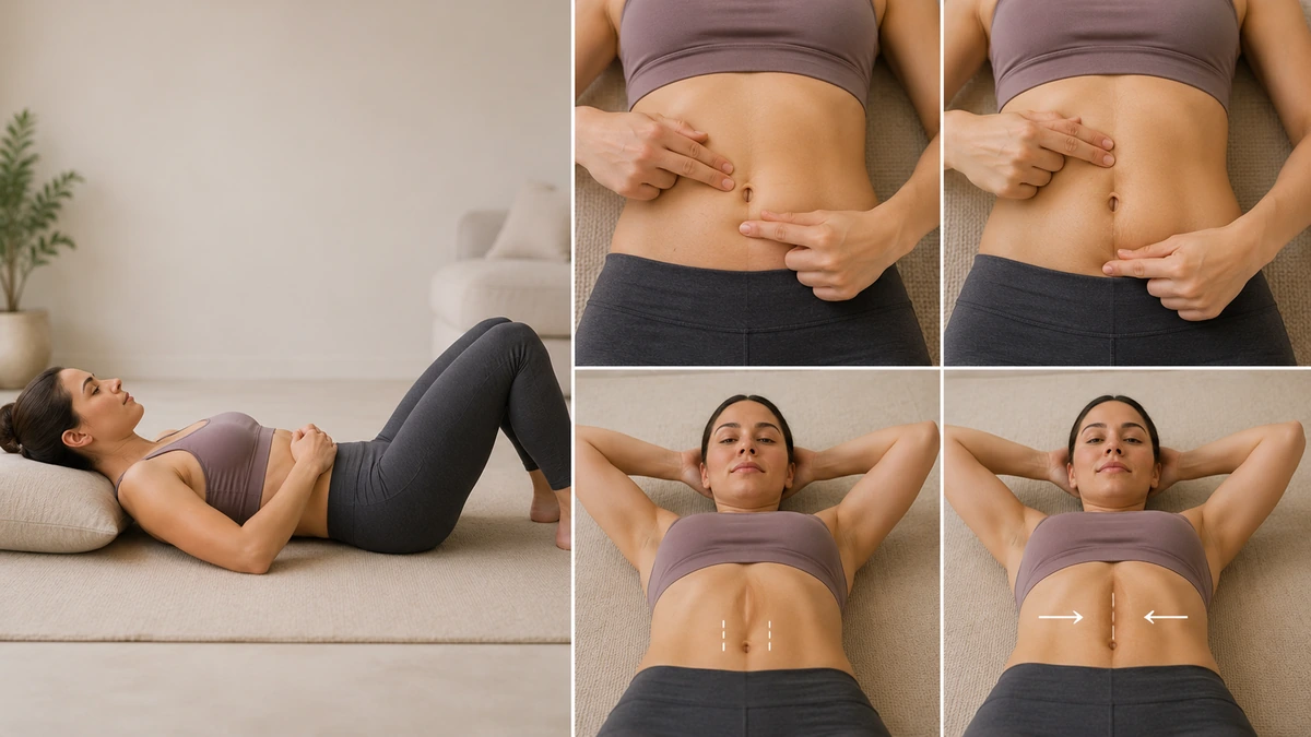
"How do I know if I have diastasis recti?" is one of the most-searched postpartum questions on the internet — around 7,800 US searches per month. The good news: you can get a workable answer in 30 seconds at home. The important caveat: self-check is a starting point, not the last word. For a complete muscle repair overview — what diastasis means for surgery candidates and what the full treatment landscape looks like — see our pillar guide. This page gives you both the exact self-check technique and the honest picture of what to do next depending on what you find.
First: What Diastasis Recti Actually Is
Diastasis recti abdominis is the separation of the two rectus abdominis ("six-pack") muscles at the midline. The two muscles are normally joined at the linea alba — a strip of connective tissue running vertically down the belly. When the linea alba is stretched (most commonly by pregnancy or significant weight change), the muscles pull apart and the gap between them widens. Full anatomical detail in our diastasis recti pillar guide.
Diastasis is extremely common. A meta-analysis of pregnancy studies indicates 30–70% of women have diastasis in the immediate postpartum period, with roughly 30% persisting past 12 months.2 It is not a rare condition — it is a normal consequence of pregnancy that resolves in some women and persists in others.
The Visual Signs
Before you touch anything, look. Stand in front of a mirror without a shirt on and examine your abdomen from the side and front. Then lie down on your back with your knees bent and lift your head slightly — as if starting a small crunch. What to watch for:
- Midline doming or ridging. A vertical ridge or dome that "pops up" along the middle of your abdomen when you engage your core. This is the single most classic visual sign. In severe diastasis, the ridge can look like a tent or ridge running from your ribs down to your pubic bone.
- "Coning" at the navel. A pointed or cone-shaped protrusion right at the belly button when you contract.
- Persistent "pooch." A soft protruding belly that does not resolve with weight loss or general exercise. Diastasis-related pooch is caused by the abdominal contents pushing forward through the weakened midline, not by fat.
- Umbilical asymmetry. The belly button may pull outward, look stretched, or appear pointed rather than round.
- Visible outline of the rectus muscles. In some cases the two separated muscles are visible as two parallel bands with a soft valley between them.
The Feel Signs
What you'll feel when you touch:
- A soft, mushy gap along the midline, between the two firm muscle bands.
- The ability to sink your fingers into the gap when the muscles are relaxed.
- The gap narrows or firms up when you engage the muscles (this is normal even in diastasis — the muscles are still pulling together somewhat).
- The tissue feels different at different points along the midline — often widest right at or just above the navel.
The Functional Signs
Symptoms you may notice in daily life:
- Lower back pain — the most common functional symptom. Diastasis reduces core stability and increases lumbar load.
- Poor core strength — feeling weaker in tasks that involve lifting, twisting, or standing for long periods.
- Urinary leakage (stress incontinence) — coughing, sneezing, or exercising causes small leaks. Diastasis frequently coexists with pelvic floor weakness.
- Feeling of core weakness or "instability" — a sense that the core isn't supporting you properly.
- Difficulty regaining pre-pregnancy shape despite weight loss and general exercise.
- Constipation or bloating — sometimes reported, related to the mechanics of intra-abdominal pressure.
- Umbilical hernia symptoms — visible navel bulge that becomes more prominent with straining. Diastasis is associated with umbilical hernia, co-occurring in 47% of abdominal-wall hernia cases per a 2025 review, particularly in severe cases.3
Not everyone with diastasis has all of these symptoms. Some women have wide gaps with minimal functional impact; others have narrower gaps with significant symptoms. The overall picture matters more than any single finding.
The At-Home Self-Check Test
The self-check is a simple test that takes about 30 seconds. It gives a workable estimate of gap width and can be repeated over time to track change. Research indicates that the finger-width method correlates well with formal ultrasound measurement when performed correctly.1
Step-by-step
- Lie on your back on a firm surface with knees bent and feet flat on the floor.
- Relax your abdomen. Do not contract yet.
- Place two fingers horizontally across your midline, right at the navel — pointing across (not up-down), fingertips toward one side of your body.
- Slowly lift your head and shoulders off the floor just enough to feel your abdominal muscles start to engage — as if beginning a small crunch. Do not do a full sit-up.
- Feel for a soft gap between the two firm muscle bands (rectus abdominis). Your fingers will sink into the gap if it's wider than 1 finger; you may need to add or remove fingers to gauge its true width.
- Estimate the width in finger-widths: 1 finger, 2 fingers, 2.5, 3, 3+.
- Also feel the depth — is the gap shallow and taut (mild) or deep and soft (more significant)?
- Repeat at two more points: about 2 inches (5 cm) above the navel, and 2 inches (5 cm) below. Diastasis width can vary — the widest point may not be at the navel.
- Record your results. Track over time if you're doing this pre- and post-pregnancy or before and after an exercise program.
How to interpret
| Gap width | Approx cm | Category | What it usually means |
|---|---|---|---|
| < 1 finger | < 1.5 cm | Normal | Not diastasis; general core work |
| 1–2 fingers | 1.5–2.5 cm | Normal-to-mild | Common postpartum; often resolves |
| 2–3 fingers | 2.5–3.5 cm | Mild diastasis | PT-guided exercise often effective |
| 3–4 fingers | 3.5–5 cm | Moderate | Exercise often helpful; may need surgery |
| > 4 fingers | > 5 cm | Severe | Surgical repair usually required |
Depth matters. A firm, taut 3-finger gap is milder than a deep, soft 3-finger gap. If you can push your fingers deep into the gap, or feel abdominal contents behind the thin tissue layer, this indicates poorer linea alba quality regardless of the width.
Limitations of the self-check
- Technique variation — even with clear instructions, self-check width estimates vary between individuals.
- Cannot assess tissue quality precisely, only estimate it.
- Cannot detect associated hernias reliably.
- Cannot assess pelvic floor function, which frequently coexists with diastasis.
For these reasons, a professional assessment adds meaningful information — but the self-check is enough to know whether you have diastasis at all and whether you need to seek that professional assessment.
Mild vs Severe — What Matters
Not all diastasis is the same. What separates mild from severe:
| Feature | Mild | Severe |
|---|---|---|
| Width | 2–3 cm (~2 fingers) | > 5 cm (> 3 fingers) |
| Tissue quality | Firm linea alba | Soft, stretched linea alba |
| Doming | Subtle with effort | Prominent, visible at rest |
| Pooch | Small, may resolve with time | Persistent, does not resolve |
| Back pain | May or may not have | Common |
| Hernia risk | Low | Elevated |
| Exercise response | Often responds well | Limited response; surgery usual |
| Time to resolution | Often 6–12 months | Rarely resolves without surgery |
When to See a Professional
See a pelvic floor physical therapist if:
- Your self-check shows a gap wider than 2 finger-widths, especially with soft/deep tissue quality.
- You have functional symptoms — back pain, urinary leakage, feeling of weakness.
- You are more than 6 months postpartum with no narrowing.
- You have tried dedicated deep-core exercises for 3 months without improvement.
- You have a visible bulge at the navel that could be a hernia.
See a board-certified plastic surgeon if:
- You have severe diastasis (> 5 cm) not responding to PT.
- You have significant associated skin laxity as well.
- You have an umbilical hernia.
- You have completed all planned pregnancies and want a definitive result.
Full information on treatment options in our how to fix diastasis recti guide, non-surgical exercise detail in our diastasis exercises guide, and surgical repair detail in our muscle repair surgery guide.
A Note for Men
Diastasis is more common in women (particularly postpartum), but men absolutely can have it. In men the cause is typically central obesity, heavy lifting without proper bracing, or genetic factors. The self-check is the same, the interpretation is the same, and the treatment framework is similar — with the important addition that weight loss is often the first and most impactful intervention. See our dedicated diastasis recti in men guide.
FAQ
How do I know if I have diastasis recti?
The most common signs are a visible bulge or "doming" along the midline of your abdomen when you sit up, lift your head off the floor, or engage your abdominal muscles; a soft or gap-like feeling between the abdominal muscles when you press with your fingers along the midline; a persistent "pooch" or protruding belly appearance that does not resolve with weight loss; and functional symptoms including lower back pain, poor core stability, urinary leakage, or a feeling of core weakness. The definitive way to confirm is the at-home self-check test (lying on your back, lifting your head slightly, feeling for a gap between the two rectus muscles at, above, and below the navel) and then confirming with a professional — a pelvic floor physical therapist can measure the inter-rectus distance accurately and assess associated tissue quality.
How do I do the diastasis recti self-check at home?
Step 1: Lie on your back with knees bent, feet flat on the floor. Step 2: Place two fingers horizontally across your midline, right at your navel. Step 3: Slowly lift your head and shoulders off the floor just enough to feel your abdominal muscles engage — as if starting a small crunch. Step 4: Feel for a soft gap between the two rectus abdominis muscle bands running vertically down your midline. Step 5: Estimate the width of the gap in finger-widths (1 finger, 2 fingers, 3+ fingers). Step 6: Repeat about 2 inches above and 2 inches below the navel — diastasis can be widest at any of these three points. Step 7: Also assess the depth and firmness of the gap — a shallow, taut gap is milder than a deep, soft one. A gap of 2 finger-widths or less that is firm is considered normal or mild; wider than 2 finger-widths, especially if soft or deep, is diastasis that warrants professional evaluation.
How big a gap is considered severe diastasis recti?
The clinical thresholds most commonly used: less than 2 cm (~1 finger) is normal or minor; 2 to 3 cm (~2 fingers) is mild diastasis; 3 to 5 cm (2 to 3 fingers) is moderate; wider than 5 cm (more than 3 fingers) is severe. The width is not the only measure that matters — depth of the gap, tissue quality of the linea alba, presence of a hernia, and functional symptoms are all relevant.1 A 3-finger gap with soft, non-supportive tissue may be more functionally significant than a wider gap with firm tissue. This is why professional assessment matters even when self-check results are informative.
Does diastasis recti cause back pain?
Yes, in many cases. The rectus abdominis muscles are part of the core stabilisation system, and when they are separated the core loses some of its ability to support the lumbar spine. Studies consistently associate diastasis with lower back pain, poor lumbo-pelvic stability, and altered posture. Not everyone with diastasis develops back pain, and not all back pain in postpartum women is caused by diastasis — but the connection is well-established and back pain is one of the most common functional symptoms that motivates women to seek diastasis-specific treatment. Non-surgical treatment (pelvic floor PT, deep-core exercises) often improves the back pain even when the gap itself does not fully close.
Can diastasis recti cause a hernia?
Yes. Diastasis and umbilical (belly button) hernias frequently coexist — a 2025 review found diastasis present in 47% of cases with a concomitant abdominal-wall hernia,3 and severe diastasis increases the risk of umbilical hernia. The mechanism: the stretched linea alba that defines diastasis is the same tissue that normally prevents abdominal contents from bulging through the navel. When this tissue is significantly weakened, a small opening can develop through which fatty tissue or bowel can protrude. Signs include a visible bulge at the navel that becomes more prominent with straining, coughing, or standing; occasional discomfort at the navel; and sometimes a small palpable soft mass. Hernias are separate from diastasis but often need to be surgically repaired at the same time as diastasis repair (during a tummy tuck).
When should I see a doctor for diastasis recti?
See a healthcare provider if: (1) your self-check shows a gap wider than 2 finger-widths, especially if it feels soft or deep; (2) you have functional symptoms — lower back pain, urinary leakage, feeling of core weakness; (3) you have a visible bulge at the navel that could be a hernia; (4) it is more than 6 months postpartum and the gap has not narrowed; (5) you have already tried dedicated deep-core exercises for 3 months with no improvement. The most useful initial referral is to a pelvic floor physical therapist — they can accurately measure the gap, assess associated pelvic floor function, and design an individualised program. If exercise is not adequate after several months of PT, a plastic surgeon consultation for potential surgical repair is the next step.