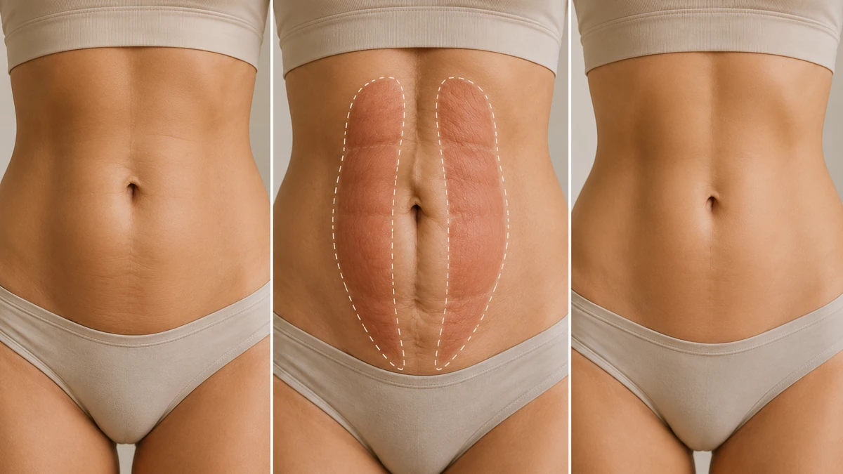
If you have ever noticed a soft mid-line bulge in your belly when you sit up out of bed, or a "dome" that pushes out when you try to do a crunch, you may be looking at diastasis recti. It is one of the most common and least dangerous abdominal-wall findings after pregnancy — and one of the most misunderstood. This guide covers what it actually is, how to check for it at home, when to worry, what exercise can and cannot fix, and when surgical repair is genuinely indicated.
Diastasis recti is not a hernia, it is not immediately dangerous, and — critically — it is not always something that needs to be fixed. The medical literature is clear that most cases improve on their own in the first year after childbirth and that structured rehabilitation, not surgery, is first-line for the ones that don't. Understanding that hierarchy is the difference between an overtreated cosmetic decision and a well-timed clinical one.
What Is Diastasis Recti?
Diastasis recti abdominis (DRA) is a widening — a "separation" — of the two halves of the rectus abdominis muscle at the linea alba, the strip of connective tissue that runs vertically down the middle of the abdomen from the breastbone to the pubis. In DRA the muscles themselves are intact; what has stretched is the fibrous mid-line seam that holds them together. Because it is a stretching of connective tissue rather than a hole in the abdominal wall, DRA is anatomically distinct from a hernia, though ventral or umbilical hernias can co-exist with it.1
A recent review defines diastasis recti as an inter-rectus distance (IRD) exceeding approximately 2 cm at any point along the linea alba at rest, though thresholds vary by measurement site and by whether the abdomen is at rest or actively contracted.1 The distinction between DRA and a true midline hernia matters: a hernia involves an actual defect through which abdominal contents can protrude, while DRA is a stretching without a defect. That is why the treatment ladders — and the risks of leaving each untreated — are different.
Who gets diastasis recti?
The classic patient is a woman in her third trimester or the early postpartum period. A large prospective cohort of 300 women followed through pregnancy and 12 months postpartum found a prevalence of about 33.1% at week 21 of pregnancy, 60% at 6 weeks postpartum, and 32.6% at 12 months.2 Those numbers matter because they show two things: DRA is extremely common in the postpartum window, and roughly half of cases resolve within a year without intervention. Older age, higher parity, elevated BMI, and diabetes are among the plausible risk factors identified in the current literature.1
DRA also occurs outside pregnancy — in men and non-pregnant women — most often in the setting of central obesity, rapid weight change, or chronic increases in intra-abdominal pressure (heavy lifting with poor bracing, chronic cough, constipation). It is less common but presents with the same anatomic finding: a widened linea alba with a mid-line bulge when the abdomen is tensed.
Symptoms & How to Check
The most common visible sign is a soft, longitudinal bulge or "dome" down the center of the belly when you try to sit up or contract the abdominal muscles. Beyond appearance, symptoms can include a feeling of core weakness, low-back or pelvic-girdle pain, and — in some patients — pelvic-floor symptoms such as urinary leakage that has not responded to standard postpartum recovery. Our symptoms and self-check guide covers each sign in detail with step-by-step diagnosis instructions.
Self-check at home
A simple palpation exam is the standard first step:
- Lie on your back on a firm surface with your knees bent and feet flat.
- Place two or three fingers horizontally just above your belly button, palm facing you.
- Slowly lift your head and shoulders off the floor, as if starting a crunch. Exhale as you lift.
- Feel for the two firm bellies of the rectus muscle and the softer gap between them. Press gently into the mid-line.
A gap wider than about two finger-widths (roughly 2 cm) at rest, a soft area you can push into, or visible doming during the lift are consistent with diastasis. Repeat the check above the belly button, at the belly button, and below it — DRA is often widest at one of the three levels. If the finding is subtle or you are trying to decide about surgery, ultrasound is the diagnostic gold standard and can quantify the inter-rectus distance at each level.3
When to see a professional
See a clinician (physician or pelvic-health physiotherapist) if the gap is wide, the bulge is painful, you have new-onset low-back or pelvic-girdle pain, or if you feel a distinct tender lump — which may indicate an associated hernia rather than DRA alone. Non-postpartum patients with a new mid-line bulge should always be assessed to rule out a hernia before assuming diastasis.
Diastasis Recti in Women vs Men
The bulk of the research on diastasis recti is postpartum-focused because pregnancy is by far the most common trigger. But DRA in men — and in women who have not been pregnant — is a genuine clinical entity that is often missed or under-diagnosed.
Postpartum women
The pattern is well characterised: the linea alba stretches under the mechanical load of a growing uterus, hormonal changes to connective tissue in late pregnancy contribute to laxity, and the mid-line often has not returned to baseline width by 6 to 12 weeks after delivery — patients who also had a Caesarean section may want to read our tummy tuck after a C-section guide for the additional timing and scar considerations. In many cases it continues to remodel over the first year. Repeated pregnancies, twin gestations, larger baby size, and higher pre-pregnancy BMI are all associated with a greater likelihood of persistent DRA.1
Diastasis recti in men
DRA in men is less studied but well recognized. It typically develops in the setting of central (visceral) obesity, where the accumulation of intra-abdominal fat progressively stretches the anterior abdominal wall. Other contributors include age-related connective-tissue changes, rapid weight change (either loss or gain), and chronic straining. The presentation is a mid-line bulge that becomes more visible when the abdomen is tensed — a curtain of soft tissue between the two rectus muscles. Non-surgical management (weight loss, core rehabilitation) is first-line; surgical repair follows the same principles as in women, though it is more often addressed alongside a hernia repair when the two co-exist.
Non-Surgical First: Exercises & Physical Therapy
The most important message on this page: exercise is first-line for persistent postpartum diastasis recti. Systematic reviews of postpartum abdominal training — pooled across dozens of studies and tens of thousands of participants — consistently find that structured programs reduce the inter-rectus distance and support core function.4 There is no single universally superior protocol; hypopressive and conventional abdominal exercises both work, and the best program is one supervised by a pelvic-health physiotherapist and tailored to your findings — our diastasis recti exercises guide covers the evidence-backed protocols in detail.
What tends to work
- Transversus abdominis activation. Gentle "drawing in" of the lower belly, held for 5 to 10 seconds and repeated. This targets the deep stabiliser that supports the linea alba from within.
- Diaphragmatic breathing with core engagement. Coordinating the diaphragm, pelvic floor, and transversus retrains the pressure system that keeps the mid-line closed under load.
- Progressive loading. Once basic control is established, movements are gradually added — bridges, modified dead-bugs, side-lying leg work — always with the mid-line watched for doming.
- Pelvic-floor integration. DRA and pelvic-floor dysfunction often co-exist; addressing them together produces better outcomes.
What to avoid — at least in the early phase
- Traditional crunches and full sit-ups. These load the rectus in a way that can widen the gap and worsen doming.
- Front-loaded planks and push-ups before core control returns. Add them back only when the mid-line stays flat under load.
- Heavy lifting with breath-holding. Learn to exhale on exertion and brace the deep core rather than pushing the abdominal wall outward.
- Anything that produces a visible dome at the mid-line. That is your live feedback signal that the load is currently too much.
How long to give rehab before considering anything else
Most guidelines recommend at least 6 months of a structured, progressive core program before concluding that rehabilitation has plateaued — and many recommend up to 12 months, particularly in the first postpartum year when spontaneous improvement is still likely.5 A physiotherapist can also identify contributing factors (posture, breathing pattern, pelvic-floor tone) that may be quietly limiting your progress.
Continue: How muscle repair fits into a tummy tuck — the full picture →When Surgery Is Considered
Surgery is not the default. The Swedish national guidelines — some of the clearest published on this topic — recommend surgical repair only when three conditions are met: at least 6 months of a structured core program has failed to produce meaningful improvement, the separation is ≥5 cm, and the patient is at least 2 years post-childbirth.5 Individual surgeons may adjust these thresholds, but they capture the general principle: rehab first, and long enough to know it has plateaued, before offering surgery. The full candidacy guide walks through BMI, health, and lifestyle thresholds that apply to any tummy-tuck decision.
Beyond the pure "gap width" criteria, surgery becomes reasonable when:
- Functional symptoms — back pain, core weakness, incontinence — persist despite competent physiotherapy.
- There is a significant skin excess or "apron" co-existing with the DRA that will not respond to exercise or weight loss (for these patients a full tummy tuck that includes muscle repair is often the appropriate answer, rather than a stand-alone repair).
- An umbilical or ventral hernia co-exists and needs surgical treatment anyway — repairing DRA at the same operation is often reasonable.
- The patient is done having children — future pregnancy can undo the repair.
The mini vs full tummy tuck decision is directly tied to diastasis: a mini leaves the upper rectus alone; if diastasis extends above the umbilicus, a full is usually needed to address it properly.
Surgical Repair Explained
The core surgical technique for diastasis recti is rectus plication: the surgeon sutures the two halves of the rectus muscle back together in the mid-line, from sternum to pubis or over whatever length is separated — our surgical repair page explains the technique, suture types, and mesh-versus-suture evidence in full. Plication has been used for decades and remains the reference technique across open, laparoscopic, and hybrid approaches, with low reported recurrence rates in modern series.6
Plication as part of a tummy tuck
For patients who also have skin excess — the typical post-pregnancy patient — plication is performed during a full abdominoplasty. After the skin is lifted, the surgeon exposes the two rectus muscles and closes the gap with a running or interrupted suture, tightening the fascia along the mid-line. When the skin is then re-draped and closed, the result is both a flatter surface (from skin removal) and a tighter core (from plication). This is the most common single surgical situation in which DRA is repaired.
Stand-alone repair (without skin removal)
When a patient has isolated DRA without significant skin excess — for example, a slim postpartum woman with a persistent bulge but no apron — a stand-alone repair may be offered. Options include an open plication through a small incision, or a minimally invasive (laparoscopic or endoscopic) approach that closes the gap without a large scar. A scoping review of surgical techniques and a comparative analysis of abdominoplasty versus minimally invasive DRA-only repair both suggest that the choice depends on the individual patient's anatomy, symptoms, and whether skin excess is present.6
Sutures alone, or with mesh?
A recent randomized clinical trial of 86 women compared plication with sutures alone against plication reinforced with a synthetic mesh, at one year post-op. The trial found no significant difference in recurrence between the two techniques, supporting the continued use of suture-only plication for most patients — a simpler, lower-cost technique with a comparable outcome at 12 months in this specific setting. Individual surgeons may still favor mesh for very wide separations or in patients with additional hernia risk.
Continue: Tummy tuck with muscle repair — how it works & who needs it →FAQ
-
What is diastasis recti?
Diastasis recti abdominis (DRA) is a separation of the two halves of the rectus abdominis muscle at the linea alba — the connective-tissue seam that runs down the middle of the abdomen. It is common in the third trimester of pregnancy and in the early postpartum period, and can also occur in men from central obesity or rapid weight change. It is not a hernia and is not immediately dangerous.1
-
How do I check for diastasis recti at home?
Lie on your back with knees bent and feet flat. Place two or three fingers just above your belly button, then slowly lift your head and shoulders like the start of a crunch. Feel for a gap between the two muscle bellies. A gap wider than about two finger-widths (approximately 2 cm) at rest — or a soft, doming area you can push into — suggests diastasis recti and is worth confirming with a physiotherapist or physician. Ultrasound is the diagnostic gold standard when the finding is unclear.3
-
Does diastasis recti go away on its own after pregnancy?
Often yes, at least in part. Prospective studies show a prevalence of about 60% at 6 weeks postpartum, falling to around 33% at 12 months.2 Most cases improve on their own in the first year without any specific treatment. Persistent separation after 12 months is less likely to resolve spontaneously and is where a structured rehabilitation program — and, in select cases, surgical repair — is considered.
-
Which exercises fix diastasis recti — and which make it worse?
Systematic reviews find that structured abdominal exercise programs — especially those that emphasise transverse abdominis activation — can reduce the inter-rectus distance in postpartum women.4 Traditional sit-ups, full crunches, planks (in the early phase), and any movement that produces visible mid-line doming can worsen the gap and should be avoided until core control returns. There is no single universally superior protocol; work with a pelvic-health physiotherapist for a program suited to your findings.
-
When is surgery considered for diastasis recti?
Surgery is considered when the separation persists beyond about 12 months postpartum despite a structured rehabilitation program, is wide (often >3–5 cm), causes functional symptoms (back pain, core weakness, incontinence unresponsive to physiotherapy), or is associated with an aesthetic apron of skin/fat that will not resolve.5 Repair is most often performed as a rectus plication during a tummy tuck, and — for isolated cases without significant skin excess — as a stand-alone laparoscopic or open plication.
-
Do men get diastasis recti?
Yes. Diastasis recti in men is less studied than the postpartum form but occurs with central obesity, rapid weight change, chronic straining (heavy lifting with poor technique), and older age. The presentation is similar — a mid-line bulge or doming when the abdomen is tensed. Non-surgical management (weight loss, core rehabilitation) is first-line; surgical repair follows the same principles as in women.